

Analysis of Skin Barrier Mechanism
Oct 30,2024
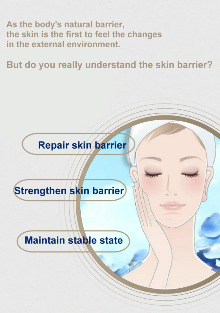
About the Skin Barrier
The skin barrier is composed of four highly interconnected layers: physical, chemical, microbial and immune barriers, which enable the skin to maintain structural integrity, protect against external aggressions, and quickly restore barrier and immune homeostasis.
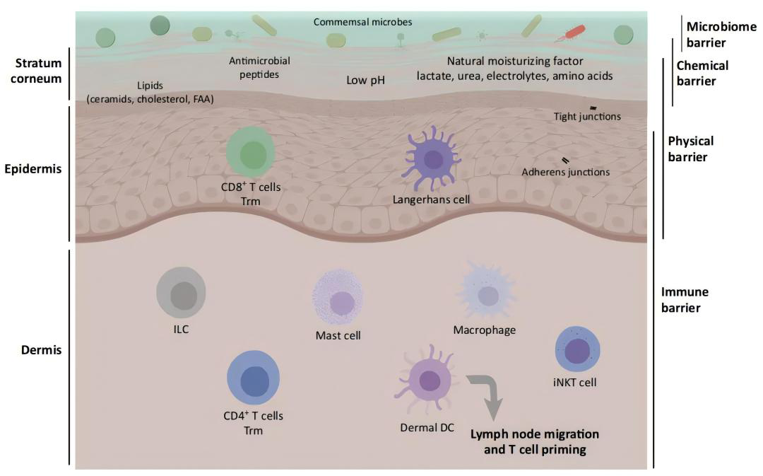
Figure 1 - Skin barrier classification and main components
01 Physical barriers
The stratum corneum (SC), the outermost layer of the epidermis, is key to maintaining a physical barrier structure that prevents transepidermal water loss (TEWL), penetration of external immunogens and microorganisms. The SC has a "brick and mortar" structure in which protein-rich corneocytes (representing the bricks, composed of maturing/ripening keratinocytes surrounded by involucrin, loricrin and keratin intermediate filaments, etc.) are interwoven in a lipid-rich extracellular matrix or lipid layer (representing the mortar, composed of ceramides, free fatty acids and cholesterol).

Figure 2 - The "brick and mortar" structure of the stratum corneum
Filaggrin (FLG) binds to intracellular keratin to promote flattening of the outer layer of the SC, and cross-linked FLG minimizes TEWL and prevents the penetration of allergens and toxins.
Ceramide, free fatty acids, and cholesterol are arranged in an intercellular lamellar structure at a ratio of 3:1:1, ensuring the three-dimensional lamellar organization of lipids, exerting hydration and protection functions. Abnormal lipid composition leads to increased TEWL and allergen penetration.
Tight junctions (TJs) limit the extracellular transport of solutes and TEWL. TJ function is also related to other components of the barrier, and the mouse claudin-1 (key TJ protein) knockout model also shows reduced FLG expression, lipid destruction, and increased inflammatory response.
02 Microbial Barrier
A diverse microbiota layer composed of commensal microorganisms and bacteria (such as Staphylococci, Corynebacterium, and Propionibacterium) colonizes the skin surface to form a microbial barrier that limits the space and nutrients for other potential pathogenic microorganisms.
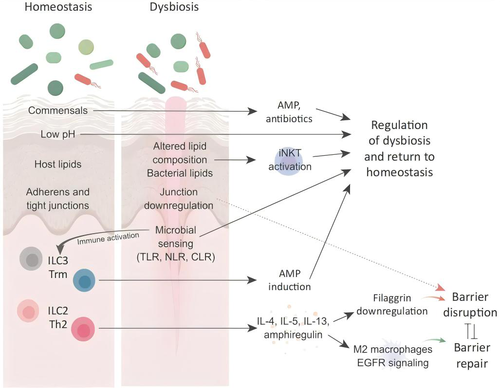
Figure 3 - A delicate balance between microbial diversity, inflammation, and barrier repair
Symbiotic microorganisms send signals to keratinocytes, inducing them to produce antimicrobial peptides (AMPs) such as defensins, S100, and LL-37, which participate in immune regulation, keratinocyte activation, and skin barrier function.
Staphylococcus epidermidis can upregulate TJ and help maintain the physical barrier, recognize symbiotic organisms through TLR-2 receptor sensitization, and prevent the host from producing immune and inflammatory responses.
Staphylococcus aureus stimulates T cells to mediate inflammatory responses, damages the skin barrier and serine proteases, activates the immune system, and stimulates the secretion of IL-4, IL-13, and IL-22.
03 Chemical Barrier
The chemical barrier consists of a series of protective molecules produced by different types of skin cells, such as AMPs, lipids, natural moisturizing factors (NMFs), and compounds that maintain acidic pH.
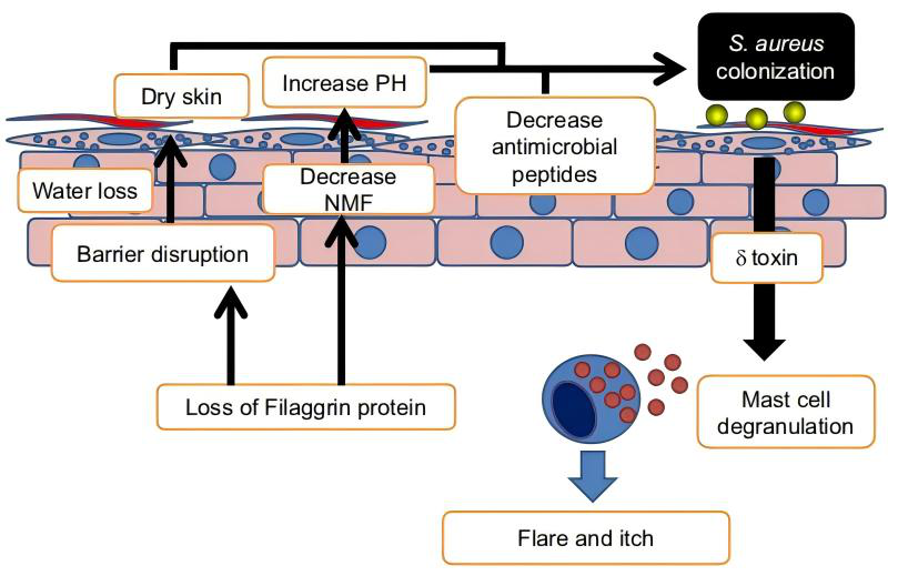
Figure 4 Chemical barrier disruption induces Staphylococcus aureus colonization
Lipids are derived from sebocytes, keratinocytes, and symbiotic microorganisms, protecting SC from TEWL, UV rays, oxidation, and pathogens.
Barrier moisturizing is mainly achieved through lipids and skin surface hygroscopic agents. FLG is decomposed by proteases to release a large amount of L-histidine, producing NMF, which absorbs moisture from the dermis into the SC.
The acidic outer membrane maintains the skin pH between 4 and 6, maintains the antimicrobial environment in the body, maintains the integrity of the barrier, and prevents bacterial imbalance. Increased skin pH disrupts the lipid bilayer structure of the SC and increases barrier permeability.
04 Immune Barrier
The immune barrier is composed of dermal dendritic cells (DCs), resident T cells, and keratinocytes. Immune cells maintain skin barrier homeostasis by recognizing barrier disruptions, communicating with commensal microorganisms, and initiating immune pathways.
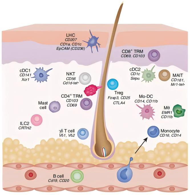
Figure 5 - Types of immune cells in human skin
Resident and recruited immune cells of both innate and adaptive cell types together constitute the skin immune barrier Innate immune cells continuously sample the external environment and selectively initiate signaling responses to barrier-damaging factors. Resident cells such as DCs, T cells, and keratinocytes can all initiate signaling responses to barrier damage.
Innate and adaptive immune responses, as well as competition from commensal organisms, prevent the growth of potential pathogenic organisms and induce inflammation.
Mutations in genes encoding FLG and TJ proteins, respectively, can weaken SC structure and increase TJ permeability, inducing the expression of cytokines such as IL-4 and IL-13. Cytokines further damage the barrier by inhibiting FLG expression and disrupting TJ.
Barrier damage and repair
The four types of barrier functions are intertwined with each other to build a solid defense line for the skin to resist the invasion of the external environment.
When the skin is subjected to external stimuli (such as physical damage, irritating chemical components, etc.), it will show dryness, desquamation, redness, etc., accompanied by discomfort such as itching and stinging, and the tolerance will decrease, becoming sensitive and prone to allergies. We call it "damaged skin barrier"
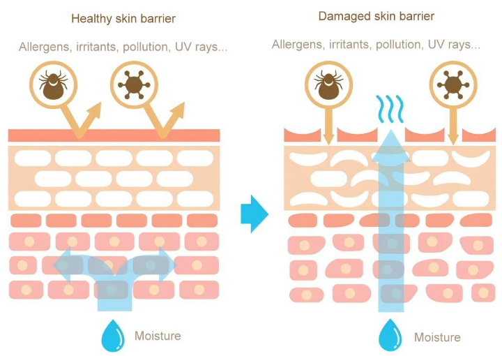
Figure 6 - Hydration of healthy and damaged skin barrier
The management of skin barrier can be done from two aspects: "repair" and "protection": first, use moisturizing/anti-inflammatory/lipid ingredients to repair and strengthen the skin barrier; second, avoid excessive cleaning, UV exposure and inferior cosmetics to keep the skin barrier stable and resist the stimulation and intrusion of the external environment with a more tenacious attitude.
A deep understanding of the structure and function of the skin barrier will allow us to prevent and manage skin problems more scientifically.

Contact
Sales Office: Bestgrand Holdings Bldg, No. 6 Nanheng West Rd, Huizhou, PRC
R&D Center: Red Maple Hi-tech Industry Park, Nanjing, PRC


